Skip to content

The University of Iowa Libraries

Skip to content
Go to
InfoHawk+
University of Iowa Libraries University of Iowa Libraries The University of Iowa The University of Iowa Libraries

Need to Know

Tag: anatomy

anatomical print from 1681 book
Mar 09 2022

John Browne | Myographia Nova | Dissection | Book of the Month from the John Martin Rare Book Room @Hardin Library

Posted on March 9, 2022 by dihrig
image of John Browne
John Browne


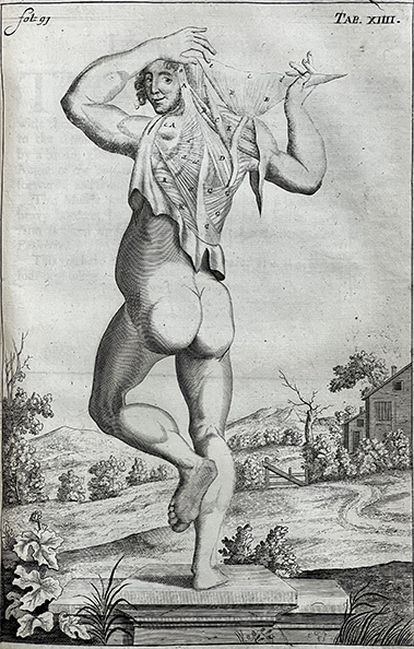
By Damien Ihrig, Curator, John Martin Rare Book Room

BROWNE, JOHN (1642-1700) Myographia nova, or, A description of all the muscles in humane body : as they arise in dissection : distributed into six lectures ; at the entrance into every of which, are demonstrated the muscles properly belonging to each lecture now in general use at the theatre in Chyurgeons-Hall, London, and illustrated with one and forty copper plates, accurately engraved after the life, with their names on the muscles, as much as can be expressed by figures, as also, with their originations, insertions, uses, and divers new observations of the authors, and other modern anatomists. Together, with an accurate and concise discourse of the heart, and its use, as also of the circulation of the blood, and the parts of which the sanguinary mass is made and framed, written by the late Dr. Lower. Printed by Thomas Milbourn for the author in 1697. 53 cm tall.

John Browne (1642-1700) was a 17th-century English surgeon and anatomist. He was royal surgeon to Charles II, James II, and William III. Under Charles II, Browne first came to observe the “King’s Evil,” or scrofula (cervical lymphadenitis), a disease related to tuberculosis that inflames and swells lymph nodes in the neck. Some thought that the touch of royalty could heal the disease, and Charles II performed many of these ceremonies. Browne vividly described the disease and ceremonies in his Adenochoiradelogia, printed in 1684.

With a recommendation from the King, Browne was appointed a surgeon at St. Thomas’s Hospital in 1683, where he trained. The Governors who oversaw the hospital, led by the Whig politician Sir Robert Clayton, disliked the freewheeling behavior of its surgical staff. The surgeons at St. Thomas felt unburdened by hospital policy because of their support by the King. However, Clayton and the Governors saw it differently and in 1691 dismissed the entire surgical staff, including Browne.

However, Browne was most well known as a serial plagiarizer and shrewd businessman. Most famously, Browne borrowed directly from the works of Giulio Casseri and William Molins for his 1675 and 1681 editions of, A compleat treatise of the muscles, as they appear in humane body, and arise in dissection. Fellow surgeon and author James Yonge heavily criticized Browne for his literary poaching.

In response, in 1697, Browne put together this book, a work that exemplified his approach to writing and selling books: he made a few tweaks to A compleat treatise… and released it under a new title. 

Browne tweaked a few things in Mygraphia nova in an attempt to distance himself from accusations of plagiarizing the works of Giulio Casseri and William Molins, most notably in the illustrations. Many of Casseri’s illustrations are somewhat whimsical, with lounging poses or those with movement, with the subject of the dissection often helpfully holding back layers of skin or muscle on their own bodies.

The illustrations shown here demonstrate such a pose and the kinds of small changes Browne made from A compleat treatise…  to Mygraphia nova. Browne’s defenders wished to credit him with the innovation of directly labeling tissues, rather than with a letter or number referenced in the text. This too, however, was a technique lifted from others, including Casseri and Molins.

Interestingly, he added a very short section on the heart as a muscle in the 1697 edition but directed the reader to an appendix that contained the well-known work of Richard Lower. He gave Lower full credit in the title, perhaps artfully exploiting Lower’s name recognition. Browne’s works were relatively popular and printed in several editions, including after his death.

picture of binding
Cover

The binding consists of contemporary leather with a deep brown color and a blind tooled (no color or gilt added) design on the front and back boards. It has raised spine bands with “Browne on the muscles” stamped in gilt over a red compartment on spine. The leather is quite cracked and the front and back hinges are loose, but it has been treated by our master conservation team and is stable for use.

Other than some browning at the front and back, more than likely due to exposure to the elements and the glue on the boards, the paper is in great condition. There are a few editorial marginalia as well.

In addition to Mygraphia nova and A compleat treatise…, we also have a facsimile of the 1697 Myografia nova and Browne’s collection of works describing glands, scrofula, and the King’s Evil touching ceremonies.

Contact curator Damien Ihrig or any others from this or other treasures: damien-ihrig@uiowa.edu or 319-335-9154 to arrange a visit in person or over Zoom.

Our copy of Mygraphia nova was generously donated by Helen Hislop, an influential physical therapist who trained and practiced at the University of Iowa.


Give a gift to the Libraries 

Posted in History of Medicine, John Martin Rare Book Room, Library ResourceTagged anatomy, John Browne, rare medical books
image of screen from anatomage table
Dec 08 2020

Introducing the Anatomage Table 7 – Fully Segmented 3D Anatomy

Posted on December 8, 2020 by Sarah Andrews

The Anatomage Table 7 is now available for use at the Hardin Library for the Health Sciences!

image of screen from anatomage table
One view from Table 7

The Table 7 is the world’s only fully-segmented real human 3D anatomy system. Users can visualize anatomy exactly as they would on a fresh cadaver. Individual structures of the body are reconstructed in accurate 3D, resulting in an unprecedented virtual dissection experience. Please email us for more information.

Features of the Anatomage Table 7

  • 4 Photorealistic cadavers
  • Physiology simulation and pathway visualization
  • 36 High-resolution regional anatomy
  • 1000+ Pathology case library

Guide to Anatomage Table 7

Schedule training on the table

head shot photo of Joe Promes, white man, beard and mustache, black suit, tartan tie
Joe Promes, Anatomage Training & Outreach Specialist

Faculty: suggestions for incorporating Table 7 into your curriculum:

  • Student Self-Study: Students are able to reserve Table 7 through the Hardin Library website for individual or group study.
  • Quizzes: Table 7 offers a quiz mode that allows instructors to create a quiz over structures of the cadaver. The Table also allows Pins to be placed on the cadaver to mimic a real-life cadaver quiz.
  • Lecture Content: Table 7 software allows for both screenshots and screen recordings. Instructors can create lecture content through the Anatomage Table 7.
  • Training for you: Joe Promes is available to give you an overview of using the table and features.

Additional information for faculty


Posted in Library Resource, Services, TechnologyTagged Anatomage Table 7, anatomy, virtual anatomy
picture of table-looks like hospital bed with giant tablet on top with skeleton showing
Sep 16 2020

NEW | Anatomage 3D Virtual Dissection Table

Posted on September 16, 2020 by Sarah Andrews
picture of table-looks like hospital bed with giant tablet on top with skeleton showing
Anatomage table

The Hardin Library now has an Anatomage virtual anatomy dissection table available.

The table is located near the service desk on the 3rd floor and is available to all users.  
 
The Anatomage table is a fully segmented real human 3D anatomy system, and the table replicates dissecting a cadaver.  Learn anatomy with an interactive, life-size touch screen experience.
 
Anatomage Information
The table was a gift from the Roy J. Carver Charitable Trust.
Posted in ResourcesTagged anatomage table, anatomy, virtual anatomy
medical images in color
Jan 08 2019

Robert Knox | Man, his structure and physiology | March 2019 Notes from the John Martin Rare Book Room @Hardin Library

Posted on January 8, 2019April 19, 2019 by Sarah Andrews

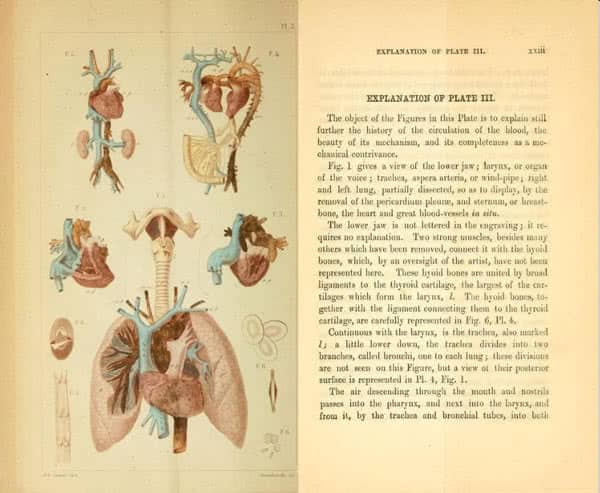
ROBERT KNOX (1791-1862). Man, his structure and physiology : popularly explained and demonstrated. 2nd ed. London ; New York: H. Bailliere, 1858.

lithograph of Robert Knox lecturing with books on stand
Robert Knox lithograph from Wellcome Trust

This popular introduction to anatomy and physiology was written by the noted – if somewhat infamous – Edinburgh anatomist Robert Knox. Knox believed that a knowledge of human structure and physiology was vital, forming the basis for a better understanding of the structure and nature of all living bodies.

He hoped that this elementary yet detailed introduction would encourage the reader to pursue further study in not only human anatomy, but also in the field of zoology. One of the best and most enthusiastic teachers of anatomy during the 19th century, Knox’s emphasis upon practical dissection led to his indiscreet and notorious association with the Edinburgh ‘resurrectionists’, Burke and Hare. When their crimes came to light, he was implicated, savagely attacked in the literature of the day, and his effigy was burnt by the populace.

Although exonerated by Burke, an influential committee of his peers accused him of acting incautiously and failing to ensure that his assistants properly vetted their cadaver suppliers. The episode haunted him and tarnished the rest of his career. He then turned his attention to ethnology and anthropology, and found some success in these fields.

You may view this book in the John Martin Rare Book Room, Hardin Library for the Health Sciences. Make a gift to the Hardin Library for the Health Sciences by donating online or setting up a recurring gift with The University of Iowa Foundation.

medical images in color
Plate III from original edition of this work from Archive.org

View a digitized copy of the original edition of this work at the Internet Archive


Posted in History of Medicine, John Martin Rare Book Room, Library Resource, ResourcesTagged anatomy, body snatching, history of medicine, Robert Knox

Categories

  • Building
  • Construction
  • Events
  • Exhibits
  • Hardin Library Staff
  • History of Medicine
  • John Martin Rare Book Room
  • Lectures
  • Library Resource
  • Nutrition
  • Plant-Based Foods
  • PubMed
  • Research Data
  • Resources
  • Scholarly Communication
  • Scholarly Impact
  • Services
  • Technology
  • Uncategorized
  • Workshops

Archives

Meta

  • Log in
  • Entries feed
  • Comments feed
  • WordPress.org
Proudly powered by WordPress | Theme: Zoia by Automattic.
University of Iowa Libraries University of Iowa Libraries The University of Iowa The University of Iowa Libraries
  • Contact the Libraries
  • Library locations & hours
  • News & Events
  • Help using the Libraries
  • Assistance for people with disabilities
  • Our diversity statement
  • Thank a Librarian
  • Web site/page feedback OR general suggestions
  • UI Libraries other links UI Libraries in the Internet Archive Use and reuse of UI Libraries web content - Creative Commons Staff SharePoint (authentication required)
  • UI Libraries on social media UI Libraries on Instagram UI Libraries on Facebook UI Libraries on Twitter UI Libraries on Pinterest UI Libraries on Tumblr UI Libraries on YouTube UI Libraries on Flickr UI Libraries blogs
  • 100 Main Library (LIB)
  • 125 West Washington St.
  • Iowa City, IA 52242-1420
  • 319-335-5299 (Service Desk)
  • ©2019 The University of Iowa
  • Give a gift to the Libraries!