from JEAN PECQUET (1622-1674). Experimenta nova anatomica, quibus incognitum hactenus chyli receptaculum, & ab eo per thoracem in ramos usque subclavios vasa lactea deteguntur. Paris: Apud Sebastianum Cramoisy et Gabrielem Cramoisy, 1651.
At the beginning of the 17th century, it was widely believed that food was converted into blood as it passed through the digestive system. The blood was then carried to the liver where it was imbued with natural spirits and passed on to the heart for distribution through the body. Since only the blood vessels were known to the anatomists of that day, it was thought that chyle, the product of digestion, was transported to the liver by the venous system of the intestines.
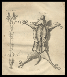
This notion was corrected by Gaspare Aselli in 1627 when, by accident, he discovered the lacteal vessels in the mesentery of a dog. He incorrectly surmised that the lacteal vessels empty their contents into the liver. It was not until 1651 that Pecquet reported his discovery of the receptaculum chyli and thoracic duct. He accurately described the lacteal veins of Aselli and showed that they terminate in the receptaculum chyli and that the thoracic duct joins the venous systems at the junction of the jugular and subclavian veins.
Joannes van Horne made the same discovery quite independently and corroborated Pecquet’s findings. Later Pecquet’s work was confirmed and extended to cover the entire lymphatic system by Olof Rudbeck (1630-1702) and Thomas Bartholin. The copperplate engraving clearly depicts the main lymphatic system both in a separate figure and in the dissected abdomen and thorax of a dog.